Left Hip Muscles Anatomy : Iliopsoas Wikipedia - The hip muscles encompass many muscles of the hip and thigh whose main function is to act on the thigh at the hip joint and stabilize the pelvis.
Left Hip Muscles Anatomy : Iliopsoas Wikipedia - The hip muscles encompass many muscles of the hip and thigh whose main function is to act on the thigh at the hip joint and stabilize the pelvis.. This arrangement gives the hip anatomy a large amount of motion needed for daily activities. The hip joint is an intricate structure including hip bones, hip articular cartilage, muscles, ligaments and tendons, and synovial fluid. Related online courses on physioplus. In conclusion, a thorough understanding of pelvic and hip anatomy is important for. The hip's unique anatomy enables it to be both extremely strong and amazingly flexible, so it can bear weight and allow for a wide range of movement.
These muscles constitute the anatomical classification known as the medial compartment of the thigh. 936 x 504 png 317 кб. These muscles are responsible for hip joint extension (backward movement). Anatomy of the muscular system. This anatomical atlas was especially designed for a specific public (radiologists, surgeons, rheumatologists and physicians specializing in musculoskeletal imaging).
Muscles Of The Hips And Thighs Human Anatomy And Physiology Lab Bsb 141 from cnx.org In order to isolate the abdominals, you need to minimize the involvement of the hip flexors and maximize the contraction of the abdominals. Pelvis and acetabulum, with muscle attachment sites. Through a simple and intuitive interface it is possible to observe every anatomical structure from any angle. Learn their anatomy efficiently and easily using kenhub's muscle anatomy and reference charts! 3 months later i got acute excrutiating pain in inguinal area. Now that you watched the video, you. Most modern anatomists define 17 of these muscles, although some additional. This 20 x 26 (51 x 66 cm) wall poster shows location of various joints and provides anterior and posterior views of the left shoulder, right hip, right knee and left elbow.
The thigh bone (femur) and the pelvis, the large bones that make up the hip joints, serve as anchors for several muscles.
I pulled some muscles on left hip hiking. Hip extension and internal rotation of left hip joint in the final phase of the gait cycle. Through a simple and intuitive interface it is possible to observe every anatomical structure from any angle. Learn their anatomy efficiently and easily using kenhub's muscle anatomy and reference charts! The hip flexors are strong, powerful muscles that can overtake the abdominal muscles in some ab exercises. Muscles of the hips and thighs | human anatomy and. Several muscles cross the front of the hip and create hip flexion, pulling the thigh and trunk toward each other, but probably the most important is the iliopsoas. Most modern anatomists define 17 of these muscles, although some additional. In human anatomy, the muscles of the hip joint are those muscles that cause movement in the hip. Rectus femoris muscle, one of the quadriceps muscles on the front of your thigh. The main functions of the neck muscles are to permit movements of the neck or head and to provide structural support of the head. Anatomical terms allow us to describe the body and body motions more precisely. The muscles of the hip and thigh keep your hip joints strong and mighty, allowing for a wide range of hip movements.
Rectus femoris muscle, one of the quadriceps muscles on the front of your thigh. Meanwhile, labral sulcus and absent labrum are normal variations in the labrum (ring of cartilage). Advanced hip flexor muscle anatomy. Through a simple and intuitive interface it is possible to observe every anatomical structure from any angle. The different anatomical areas of the gluteal region:
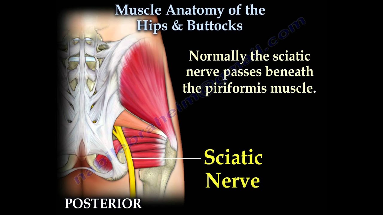
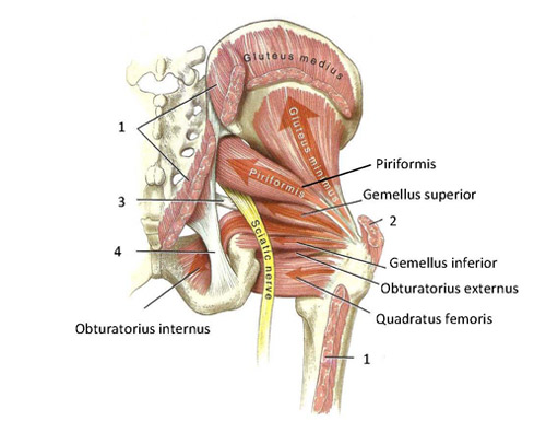
Muscle Anatomy Of The Hips Buttocks Everything You Need To Know Dr Nabil Ebraheim Youtube from i.ytimg.com The hip joint is an intricate structure including hip bones, hip articular cartilage, muscles, ligaments and tendons, and synovial fluid. Pelvis and acetabulum, with muscle attachment sites. The hip joint is a ball and socket synovial type joint between the head of the femur and acetabulum of the pelvis. In order to isolate the abdominals, you need to minimize the involvement of the hip flexors and maximize the contraction of the abdominals. Anatomy of a human body we study anatomy. Microscopic anatomy of skeletal muscle. In conclusion, a thorough understanding of pelvic and hip anatomy is important for. The different anatomical areas of the gluteal region:
Several muscles cross the front of the hip and create hip flexion, pulling the thigh and trunk toward each other, but probably the most important is the iliopsoas.
Several muscles cross the front of the hip and create hip flexion, pulling the thigh and trunk toward each other, but probably the most important is the iliopsoas. Through a simple and intuitive interface it is possible to observe every anatomical structure from any angle. I pulled some muscles on left hip hiking. Highly detailed 3d models, with textures up to 4k resolution, enable to examine the shape of each. Now that you watched the video, you. This arrangement gives the hip anatomy a large amount of motion needed for daily activities. Back muscles of the hip. In conclusion, a thorough understanding of pelvic and hip anatomy is important for. The psoas major muscle (usually shortened to just the psoas muscle) is one of the muscles of the posterior abdominal wall and lies not in the retroperitoneum but posterior to it, in the iliopsoas compartment. 1 hip anatomy, function and common problems. Most modern anatomists define 17 of these muscles, although some additional. The muscles of the neck can be divided into groups according to their location. A bursa that sometimes causes problems in the hip is sandwiched between the bump on the outer hip (the greater trochanter) and the muscles and tendons that cross over the bump.
Advanced hip flexor muscle anatomy. Related online courses on physioplus. 3 months later i got acute excrutiating pain in inguinal area. If you know all the hip flexor names and bones they attach to, that's an awesome accomplishment! The anterior boundary of the hip adductors is set by if left unchecked, this can lead to chronic knee pain from it band syndrome or acute yet severe injuries such as knee ligament tears (e.g.
Functional Anatomy Of The Small Pelvic And Hip Muscles Completed Institute Of Basic Medical Sciences from www.med.uio.no Most modern anatomists define 17 of these muscles, although some additional. Advanced hip flexor muscle anatomy. Anatomy of a human body we study anatomy. The muscles of the neck can be divided into groups according to their location. The main functions of the neck muscles are to permit movements of the neck or head and to provide structural support of the head. 1 hip anatomy, function and common problems. Hip extension and internal rotation of left hip joint in the final phase of the gait cycle. Each muscle below has the bones in bold for intermediate learners and the specific bony landmarks for advanced learners.
Related online courses on physioplus.
Learn their anatomy efficiently and easily using kenhub's muscle anatomy and reference charts! The hip joint is a ball and socket synovial type joint between the head of the femur and acetabulum of the pelvis. Meanwhile, labral sulcus and absent labrum are normal variations in the labrum (ring of cartilage). In human anatomy, the muscles of the hip joint are those muscles that cause movement in the hip. The hip joint is the articulation of the pelvis with the femur, which connects the axial skeleton with the lower extremity. There are a lot of muscles of the hip and thigh. 3 months later i got acute excrutiating pain in inguinal area. The hip's essential muscles are the sartorius, rectus femoris, gluteus minimus and medius, iliopsoas, adductors, and hamstrings. The different anatomical areas of the gluteal region: This arrangement gives the hip anatomy a large amount of motion needed for daily activities. The muscles of the neck can be divided into groups according to their location. Back muscles of the hip. Most modern anatomists define 17 of these muscles, although some additional muscles may sometimes be considered.


0 Komentar